The degree of background parenchymal enhancement (BPE) affects how well breast MRI performs, according to research published May 27 in Radiology.
Minimal or mild BPE is tied to higher sensitivity and specificity, while moderate or marked BPE is linked to lower diagnostic performance, suggest findings by Sonja Bechyna, MD, and Pascal Baltzer, MD, from the Medical University of Vienna in Austria.
“The degree of BPE in MRI reports may prompt follow-up strategies and additional imaging to prevent missed diagnoses and unnecessary biopsies,” Bechyna and Baltzer wrote.
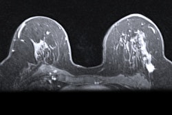
Breast MRI is the most sensitive modality for detecting breast cancer. However, its diagnostic performance can be negatively affected by BPE, where benign breast tissue also enhances after a contrast agent is administered. This creates a masking effect for malignant tumors. And the available literature is conflicting when it comes to how BPE degree affects diagnostic evaluation via breast MRI.
Bechyna and Baltzer determined whether the presence of moderate or marked BPE in women undergoing breast MRI affects the modality’s diagnostic performance. They also compared these results to those regarding minimal or mild BPE.
 RSNA
RSNA
The research duo performed a systematic review of studies published until May 2024. For the work, the team included eight studies containing data from 6,044 women with an average age of 52 years. The investigators included studies that provided raw data to extract or calculate true-positive, false-positive, true-negative, and false-negative results.
The authors reported significant associations between minimal or mild BPE and better diagnostic performance by breast MRI compared to exams with moderate or marked BPE.
Comparison between MRI exams with minimal or mild BPE, exams with moderate or marked BPE | |||
Measure | Minimal or mild BPE | Moderate or marked BPE | p-value |
Sensitivity | 95.3% | 84.1% | 0.02 |
Specificity | 89% | 78.7% | 0.02 |
Area under the curve (AUC) | 0.95 | 0.91 | 0.007 |
Also, moderate or marked BPE and publication year in cases of moderate or marked BPE were covariates influencing the diagnostic odds ratio (OR), the duo found. Finally, moderate or marked BPE remained an independent predictor of the diagnostic performance on multivariable analysis (OR, -1.33; p = 0.002).
Bechyna and Baltzer called for future studies to standardize BPE measurement similar to how fibroglandular tissue is, as well as to explore how offering different follow-up protocols or additional imaging could minimize interval cancer risk.
“Evaluation of reduced doses of MRI contrast agent could reduce the degree of BPE while maintaining lesion conspicuity,” they wrote. “Additionally, the characteristics of BPE, not just its degree, should be investigated for their diagnostic impact, as no previous studies have addressed this aspect.”
In an accompanying editorial, Manisha Bahl, MD, from Harvard Medical School in Boston, wrote that these results should lead to more research about how BPE is characterized, reported, and integrated into imaging protocols.
“Continued development of automated tools, in addition to advanced imaging techniques, will be critical to refining our approach,” she wrote. “As our understanding of BPE evolves, so too will our ability to improve the precision and clinical utility of breast MRI.”
The full study can be found here.