A novel knee cartilage relaxometry MR imaging technique provides a reliable and reproducible way to assess cartilage degeneration and could pave the way for better early detection, monitoring, and outcomes related to osteoarthritis, according to findings presented May 15 at the International Society for Magnetic Resonance in Medicine (ISMRM) 2025 annual meeting.
The technique demonstrated excellent agreement between the accelerated high-resolution imaging and the reference, noted the team working on the multivendor, multisite project that included the Cleveland Clinic, New York State University at Buffalo, University of California, San Francisco, Albert Einstein College of Medicine, and the University of Kentucky.
In a continuation of a previous project, the team said they have harmonized their model to minimize intrasite, intervendor variability to less than 5%. The work addresses needs around early detection of osteoarthritis (OA), such as a sensitive tool to monitor disease progression and treatment response.
"MRI [T1-rho] T1ρ and T2 relaxation times have been suggested as promising biomarkers for early detection of OA; however, this technique is limited to relatively low resolution," explained Zhiyuan Zhang from the Lerner Research Institute's Program of Advanced Musculoskeletal Imaging (PAMI) at the Cleveland Clinic in Ohio.
To achieve high resolution, low scan time is needed, Zhang added, saying that the team's previous work showed that compressed sensing reconstruction not only reduced scan time but preserved image quality and quantitative accuracy.
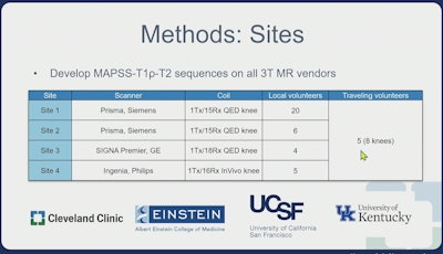
Now evaluating their technique in a multisite, multivendor study, Zhang and colleagues used a 3D MAPSS [magnetization-prepared angle-modulated partitioned k-space spoiled gradient echo snapshots] sequence on three 3-tesla MR scanners: Prisma by Siemens Healthineers with a 15-channel knee coil, Signa Premier by GE HealthCare with an 18-channel knee coil, and Ingenia by Philips with a 16-channel knee coil.
 Zhiyuan Zhang, ISMRM 2025
In all, 33 volunteers underwent scanning at four sites around the U.S., including one study volunteer who, in a unique aspect according to Zhang, traveled around the country undergoing scans of their knee at all four sites.
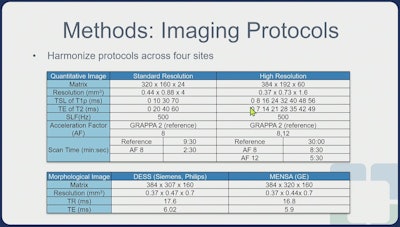
Zhang noted harmonized protocols for the study as an important aspect to achieve high reproducibility.
 Zhiyuan Zhang, ISMRM 2025
Reference images were reconstructed using GRAPPA [generalized autocalibrating partially parallel acquisitions] and coil sensitivity maps to generate coil-combined images, the team noted. Accelerated images were reconstructed using spatiotemporal finite difference transform.
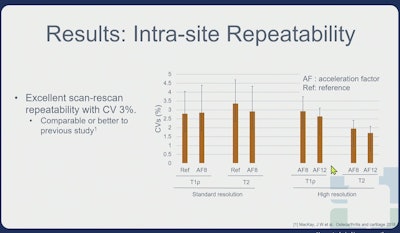
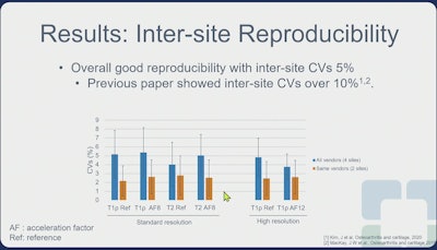
Intrasite coefficients of variation (CVs) were calculated based on the mean compartment values for comparisons between reference and accelerated images, scan and rescan, and standard versus high resolutions, according to the group. Intersite CVs were also calculated based on the mean compartment values.
Overall, the CV was 4.5 for the standard resolution and 2.6 for the high resolution, Zhang noted.
For their statistical analysis, the team evaluated:
- Agreement of T1ρ and T2 quantifications between the reference and accelerated images.
- Repeatability of T1ρ and T2 quantifications using the scan-rescan images
- Intersite variations
 Zhiyuan Zhang, ISMRM 2025
Especially with high resolution with high acceleration factor (AF) of 12, the technique still preserved very good repeatability which shows robustness at higher acceleration imaging, Zhang said.
 Zhiyuan Zhang, ISMRM 2025
Zhang also noted "good reproducibility" with a CV of 5%, which is significantly improved from the previous study, which reported CV of 10%.
Notably, the technique reduced scan time to 2 minutes, 30 seconds (approximately 70% reduction) for standard resolution imaging and to 5 minutes, 30 seconds (approximately 85% reduction) at high resolution, Zhang said.
"This technique holds promise for improving the early detection and monitoring of cartilage degeneration, ultimately leading to better patient outcome," Zhang concluded.
Check out AuntMinnie’s full coverage of ISMRM 2025 here.

