An automated brain volumetry model for MRI shows promising accuracy in distinguishing frontotemporal dementia (FTD) from Alzheimer's disease and normal cognition, according to study results to be presented May 15 at the ISMRM meeting.
Seung Hyun Lee and colleagues from Yonsei University’s College of Medicine in Seoul noted that the atrophy patterns of FTD are variable and may overlap with those seen in conditions such as Alzheimer’s disease, which can make diagnosis of the condition and distinguishing it from other neurodegenerative conditions challenging.
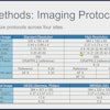
In a retrospective study, 759 patients who had undergone 3D T1-weighted MRI on 3-tesla scanners and had available Mini-Mental State Examination (MMSE) scores were divided into three groups: FTD, Alzheimer’s disease, and cognitively normal. The research team used Vuno Med DeepBrain software to measure key volumetric features of the brain, including the frontal lobes, insula, cingulate cortex, and subcortical gray matter.
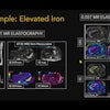
 Workflow for FTD, AD, and CN classification using automated brain volumetry. Graphic and caption courtesy of Seung Hyun Lee and the ISMRM.
The investigators identified regions with significant volume differences between FTD and Alzheimer’s disease and significant volumetric differences between FTD and Alzheimer’s disease, particularly in the frontal lobes, insula, cingulate cortex, and subcortical gray matter. They used these regions, along with age and MMSE scores, as key input variables for the model.
Using the training dataset, the model's performance demonstrated an accuracy of 89.3%, precision of 90.7%, and recall of 86.7%. The internal validation set demonstrated an accuracy of 92%, precision of 88.9%, and recall of 88.5%. External validation with an independent dataset yielded an accuracy of 84.6%, precision of 77.2%, and recall of 72.3%.
Based on these results, the tool shows promise for diagnosing FTD, the authors said.
Check out AuntMinnie.com’s full coverage of ISMRM 2025 here.