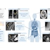
Over the last half-century, several eureka moments have powered advances that have made MRIs an indispensable diagnostic and decision-making tool across nearly every area of medicine. For example, the discovery of gradient coils allowed for faster image acquisition. Stronger magnetic fields led to improved image quality. Advances in superconducting magnets improved the signal-to-noise ratio.
 Roland Rott.
However, while advances in MRI technology are transforming patient care, the growing demand for scans is also fueling a radiology burnout crisis, staffing shortages, and long patient wait times in many parts of the country. Today, in the U.S., an unprecedented 40 million MRI scans are being performed each year. The versatility of modern MRI -- combined with an aging population and our healthcare system’s growing embrace of preventive medicine -- is why the work of a radiologist is never done.
Clear imaging has become critical to guiding therapeutic decision-making, increasing the pressure on radiologists to deploy technology and minimize issues related to patient movement on the table. Patients who are claustrophobic, can’t stay still, or are unable to hold their breath for required intervals produce “noise” that can render images diagnostically unreliable -- potentially with life-or-death consequences. Fortunately, AI has matured in the medical imaging field just in time to help understaffed, overworked radiology practices address these problems.
The following are four ways AI is improving the modern MRI to help clinicians meet the overwhelming demands on their time.
Reducing patient wait times
Aging populations and higher rates of chronic conditions have driven up demand for MRI scans. Long wait times for MRI scans are not unusual in many hospitals and healthcare systems, with wait times of 65 to 105 days, according to two studies in Canada. Shortages of radiologists or imaging technicians can also create delays in reading and interpreting image results.
Long wait times for MRI scans can delay diagnoses and treatment decisions with serious health repercussions for patients with progressive diseases. With cancer, a delayed diagnosis can give aggressive cancers time to metastasize and harm a patient’s prognosis. For patients with neurological conditions, treatment delays can lead to irreversible organ damage. Delayed MRIs for acute injuries can lead to potentially chronic issues or even disability.
This is where AI can help. With so much uncurated medical data waiting to be examined and many radiologists overloaded on cases, AI has the ability to analyze MRI scans to flag potentially serious cases so that patients with critical conditions are prioritized.
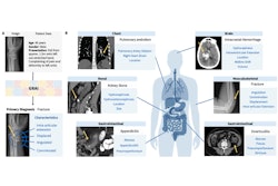
AI algorithms may be calibrated to help identify abnormalities on scans such as tumors, fractures, or signs of disease. AI systems also have the potential to act as a kind of “intelligent assistant” and offer second opinions on interpreting scans, which can be particularly helpful in rural hospitals that may have only one radiologist on duty.
Addressing radiologist burnout
Many clinicians are burnt out and actively considering leaving the healthcare industry. A RSNA survey of 13,000 radiologists in 2022 found that 49% reported burnout, with the top cause of burnout (60%) being excessive bureaucratic tasks. One-third reported excessive work hours and lack of autonomy over their life, while 28% reported frustrations related to the use of medical records.
AI-powered MRI technology has emerged as a lifeline to a growing number of short-staffed radiology departments, enabling the clinical staff to diagnose medical problems more quickly and with greater confidence. For example, sophisticated image reconstruction functionality can correct for patient movement and alleviate some of the additional burden from time-constrained clinicians to see through background noise and artifacts on MRI scans.
AI can also help improve workflow efficiency so clinicians can spend more time with patients and focus more on the right care pathway. AI orchestration is an enterprise capability, designed to enable healthcare providers to access a curated selection of clinical imaging applications with minimal effort. It provides a single contact for sourcing a variety of clinical applications and a validated, user-friendly process for integrating AI into radiology reading workflows. This supports clinicians, and it may result in improved efficiency, accuracy, and quality of radiology services.
Solving the cardiac breath hold
Cardiac MRI is used for assessing the structure and function of the heart in cardiovascular disease; however, its adoption globally remains relatively low, in part due to lengthy scan times.
Conventional cardiac MRI is too slow to capture all the frames needed across a heartbeat, so for decades, clinicians imaged and stitched together portions of data across multiple scans. This requires the patient to hold their breath to avoid motion artifacts. This process is time-consuming, prone to image quality degradation, and exhausting for patients.
Deep-learning reconstruction has emerged to address the growing demand for fast, high-quality cardiac MRI scans. This AI technology can dramatically accelerate image acquisition, with the ability to scan up to 12 times faster compared to conventional methods, enabling rapid cardiac functional imaging in as fast as a single heartbeat, matching the speed of MRI to the speed of physiology.
This advancement minimizes or removes the need for repetitive patient breath-holds, simplifying procedures and expanding the pool of patients eligible for cardiac MRI to include arrhythmic patients and those with difficulty holding their breath or the inability to remain still.
Ending the image quality versus speed tradeoff
AI is also easing a fundamental trade-off in MRI imaging: scan time versus image quality. A high-quality image -- necessary for a confident diagnosis -- demands a higher signal-to-noise ratio (SNR) and sharp visibility of body structures. Just as a photographer might leave the camera’s shutter open for a few extra moments in a dark environment to let in more light, radiologists know that the longer the scan, the better the SNR and level of detail.
AI makes it possible to have both high-quality images and shorter scan times, using deep-learning algorithms that reduce noise and improve resolution. By operating on raw data acquired during the scan, AI techniques can reproduce high SNR and sharpness with less raw data. That means scan times can be shorter with less opportunity for patient motion.
Using denoising deep-learning technology, AI is improving MRI results for patients who have trouble holding still -- such as children, those with neurodegenerative disease, and metastatic cancer patients who can’t hold their breath long enough for a traditional high-resolution MRI.
These advances are only the beginning. Each day, we are seeing new progress in the ways AI can be leveraged by clinicians to improve medical care for patients and clinicians across therapeutic areas. From oncology to neurological disorders, AI and the latest imaging technologies are going to change the way diseases are prevented, diagnosed, and treated.
Roland Rott is president and CEO of Imaging, GE HealthCare (GEHC).
The comments and observations expressed herein do not necessarily reflect the opinions of AuntMinnie.com, nor should they be construed as an endorsement or admonishment of any particular vendor, analyst, industry consultant, or consulting group.