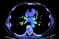
An AI algorithm can improve radiologists' identification of incidental pulmonary emboli (IPE) on CT imaging, researchers have found.
The results suggest that AI could serve as an effective "second look" tool for this indication, wrote a team led by Ronald Kuzo, MD, of the Mayo Clinic in Rochester, MN. The group's findings were published August 4 in the Journal of Thrombosis and Haemostasis.
"The AI algorithm we used has good sensitivity with a reasonably low false positive rate and can be used as a second look to improve detection of emboli that may have otherwise been missed," Kuzo and colleagues noted.
IPEs are sometimes identified on routine, contrast-enhanced CT imaging performed for other reasons than suspicion of pulmonary embolism (PE), the group explained. But because these exams are being conducted for other indications, radiologists may miss IPE. To mitigate this, using AI as an additional diagnostic tool could prove useful, Kuzo et al wrote.
The investigators tested this hypothesis via a study that used a commercially available AI algorithm (Aidoc BriefCase for iPE Triage) to review 14,453 contrast-enhanced outpatient CT chest, abdomen, and pelvis exams in 9,171 patients where PE was not suspected; they also used a natural language processing (SimpleNLP) algorithm to search radiology reports for any IPE. Thoracic radiologists reviewed all cases read as positive by AI or natural language processing to confirm incidental pulmonary emboli and evaluate "the most proximal level of clot and overall clot burden," the group explained. There were 1,400 cases read as negative by both the initial radiologist and AI that were reviewed again to assess for any IPE.
The researchers reported the following:
- Radiologists detected 218 incidental pulmonary emboli, and AI detected 224 (36 of which were missed by the radiologists). "Those missed emboli were small and distal, and the overall clot burden was low," the team wrote.
- AI missed 30 cases of IPE detected by the radiologist and had 94 false positives.
- For 36 incidental pulmonary emboli missed by the radiologist, median clot burden was one, and 19 were solitary segmental or subsegmental.
- For 30 incidental pulmonary emboli missed by AI, one case had large central emboli. The others were small, with 23 solitary subsegmental emboli.
- Radiologist re-review of 1,400 exams interpreted as negative found eight additional cases of IPE.
- The most common location of emboli missed by radiologist readers was the posterior basal segment of the right lower lobe (32%).
They also found that the specificity for identifying IPE by radiologists alone was 65.1%, for the AI algorithm, 66.9%, and for a combination of the two, 75.8%.
The results are promising when it comes to using AI for this indication, but more work is needed, according to the team.
"Compared with radiologists, AI had similar sensitivity but reduced positive predictive value," it concluded. "Our experience indicates that the AI tool is not ready to be used autonomously without human oversight, but a human observer plus AI is better than either alone for detection of incidental pulmonary emboli."
The complete study can be found here.


