An object on a conventional digital mammogram was highly suspicious for malignancy. The structure was seen in both the MLO and CC views, and persisted on the multiple diagnostic mammograms. The patient underwent biopsy and the structure was benign.
If you look at the corresponding tomosynthesis images, you see nothing malignant. The structure seen in the conventional mammogram was the result of overlapping tissues. Diagnosis was given as superimposed parenchyma.
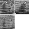
At another height in the breast, a series of calcifications can be seen in the tomosynthesis cine loop. Because of the 3D nature of the tomosynthesis images, these are known to be within a few millimeters of the skin’s surface.
This image is from a technology that is a work-in-progress and that does not have regulatory approval in certain global markets.
False positive for mammography resolved by tomosynthesis
Aug 21, 2006
Latest in Breast

Study highlights risk-based breast cancer screening; ACR responds
December 12, 2025
Radiologists can designate nonmass lesions on breast ultrasound
December 9, 2025
ACR talks BI-RADS version 2025 with AuntMinnie
December 8, 2025

DeepHealth unveils DeepHealth Breast Suite
December 2, 2025