Gadoxetate-enhanced MRI improves screening sensitivity for hepatocellular carcinoma (HCC) compared to ultrasound, according to a study published February 10 in Radiology.
"Most participants [in our study] undergoing HCC screening had excellent MRI quality even when ultrasound was limited," wrote a team led by Sara Lewis, MD, of Icahn School of Medicine at Mount Sinai in New York City.
HCC screening and surveillance are typically conducted with ultrasound, but previous research has suggested that gadoxetate-enhanced MRI may improve sensitivity. In any case, identifying factors that may affect the image quality of the two modalities is important, but "comparative data between ultrasound and MRI are lacking," according to the group.
To address this knowledge gap, Lewis and colleagues compared gadoxetate-enhanced MRI and ultrasound image quality in patients with cirrhosis undergoing HCC screening via a secondary analysis of a two-center North American HCC screening study conducted between September 2020 and May 2023 that included 245 participants with cirrhosis who underwent both gadoxetate-enhanced MRI and liver ultrasound.
Two radiologists evaluated the MRI scans for dynamic phase motion, hepatobiliary phase liver uptake, and diffusion-weighted imaging quality, and summarized their findings into the following scores: MR-A (no or minimal image limitations), MR-B (moderate image limitations), and MR-C (severe image limitations). They also scored ultrasound image quality, using the Liver Imaging Reporting and Data System (US-A: no or minimal limitations; US-B: moderate limitations; and US-C: severe limitations).
Comparison of contrast-enhanced MRI to ultrasound for HCC screening sensitivity | |
Score | Percent sensitivity |
MRI | |
| MR-A | 80.4% |
| MR-B | 18.4% |
| MR-C | 1.2% |
Ultrasound | |
| US-A | 24.2% |
| US-B | 61.7% |
| US-C | 14.1% |
The investigators reported that the proportion of exam scores with no or minimal imaging limitations was higher for MRI than ultrasound in all participants and in both sets of scores (p < 0.001).
They also found, however, that obesity (body mass index equal to or more than 30) reduced image quality in both modalities, with MRI demonstrating a univariable odds ratio [OR] of 4.2 (p = 0.01) and ultrasound demonstrating a univariable OR of 2.5 (p = 0.04) -- and that a Child-Pugh score of B and/or C reduced MRI quality at univariable (OR, 2.6; p = 0.03) and multivariable (adjusted OR, 3.95; p = 0.006) analysis. (The Child-Pugh scoring system assesses the prognosis and severity of liver disease and cirrhosis via a scoring range from five to 15; scores are divided into Class A [5 to 6 points, mild], B [7 to 9 points, moderate], or C [10 to 15 points, severe]).
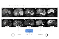
![Images in a 64-year-old obese man (body mass index, 35.2, calculated as weight in kilograms divided by height in meters squared) with metabolic dysfunction–associated steatotic liver disease who underwent hepatocellular carcinoma screening demonstrate discordant quality between MRI and ultrasound (US). (A) Poor liver visualization was noted at US (severe limitations [US-C]), with severe beam attenuation and shadowing, resulting in most of the liver not being visualized. (B, C) The quality of the MRI scans was excellent, with (B) minimal motion in the arterial phase on the postcontrast T1-weighted image and (C) no limitations on the delayed hepatobiliary phase image in the axial plane. Images and caption courtesy of the RSNA.](https://img.auntminnie.com/mindful/smg/workspaces/default/uploads/2026/02/2026-02-10-radiology-lewis-figure2.CM73B4geqo.jpg?auto=format%2Ccompress&dpr=2&fit=max&q=70&w=700) Images in a 64-year-old obese man (body mass index, 35.2, calculated as weight in kilograms divided by height in meters squared) with metabolic dysfunction–associated steatotic liver disease who underwent hepatocellular carcinoma screening demonstrate discordant quality between MRI and ultrasound (US). (A) Poor liver visualization was noted at US (severe limitations [US-C]), with severe beam attenuation and shadowing, resulting in most of the liver not being visualized. (B, C) The quality of the MRI scans was excellent, with (B) minimal motion in the arterial phase on the postcontrast T1-weighted image and (C) no limitations on the delayed hepatobiliary phase image in the axial plane. Images and caption courtesy of the RSNA.
Images in a 64-year-old obese man (body mass index, 35.2, calculated as weight in kilograms divided by height in meters squared) with metabolic dysfunction–associated steatotic liver disease who underwent hepatocellular carcinoma screening demonstrate discordant quality between MRI and ultrasound (US). (A) Poor liver visualization was noted at US (severe limitations [US-C]), with severe beam attenuation and shadowing, resulting in most of the liver not being visualized. (B, C) The quality of the MRI scans was excellent, with (B) minimal motion in the arterial phase on the postcontrast T1-weighted image and (C) no limitations on the delayed hepatobiliary phase image in the axial plane. Images and caption courtesy of the RSNA.
These scoring systems "are intended to serve as a starting point for future investigations," the group wrote.
"We recognize that quality score A -- no or minimal limitations -- has different practical implications for each modality, as ultrasound remains a screening modality whereas MRI is still considered a diagnostic modality … Quality and adequacy of an examination will depend on the clinical task (i.e., surveillance or diagnosis), the sequences included, and how the sequences were weighted."
Access the full study here.