

Biomarkers found on MRI, CT, and ultrasound can help clinicians identify Crohn disease small bowel strictures, researchers have reported in a literature review published October 16 in Radiographics.
The findings could translate into better patient care, since assessing these "penetrating and obstructing complications" is crucial for determining the best type of treatment for Crohn disease patients, noted a team led by Safa Hoodeshenas, MD, of the Mayo Clinic in Rochester, MN.
"Of particular importance is the development of accurate and dependable biomarkers for intestinal strictures, which are essential for identifying patients likely to respond to antifibrotic therapies," Hoodeshenas and colleagues wrote. "These biomarkers will be vital for improving the management of Crohn disease by enabling precise patient stratification and rigorous monitoring in clinical trials of innovative antifibrotic drugs."
Crohn disease-related strictures -- that is, narrowed segments of intestine caused by chronic inflammation and scarring -- are a serious complication of small bowel Crohn disease and can result in repeated emergency room visits or hospitalizations, endoscopic procedures, surgery, cumulative bowel damage, and disability, the authors explained. Depending on their type, strictures can be treated by medical therapy, endoscopic balloon dilation, or surgery; 40% of patients with the disease will develop strictures, and of those, 80% will require surgery.
Hoodeshenas and colleagues conducted a literature review regarding the use of various imaging modalities to identify fibrostenosing Crohn disease-related strictures. They noted that previous studies have shown that "maximum associated small bowel dilation, stricture length, and the development of new strictures are significantly associated with subsequent surgery in patients with fibrostenosing Crohn disease," and listed characteristics that could help identify those at high risk for subsequent surgery, including fibrosis, luminal narrowing, muscle hypertrophy and hyperplasia, and stiffness.
The team then described the benefits of various imaging modalities for identifying stricture biomarkers:
- MRI. "Quantitative biomarkers for CD strictures have been developed and are in various stages of validation," the group wrote. MRI techniques to identify Crohn disease biomarkers include MRI-magnetization transfer, delayed gadolinium enhancement, and diffusion-weighted imaging.
- MRI T1 mapping. "Intestinal T1 mapping is being investigated as a biomarker for assessing intestinal inflammation and fibrosis," the authors noted.
- Ultrasound. Ultrasound-based studies "generally define a stricture when bowel wall thickening exceeds 3 mm, the lumen is narrowed less than 1 cm, and there is upstream dilation greater than 2.5 cm," the team explained.
- Elastography, both ultrasound and MRI. "Tissue stiffness as a biomarker for fibrosis can be quantified by elastography techniques at MRI and ultrasound."
- PET/MR elastography (MRE). "The promise of PET/MRE for assessing Crohn disease-related strictures lies in its potential for improving the ability to distinguish between inflammation and fibrosis," Hoodeshenas and colleagues wrote.
- Fibroblast activation protein inhibitor (FAPI) PET. "Gallium-68 FAPI PET targets fibroblast activation protein, which acts as a biomarker for activated fibroblasts," they explained.
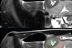
 The Stenosis Therapy and Anti-Fibrotic Research (STAR) Consortium definition of a single stricture. (A) Coronal MR image and illustration show a single bowel segment with continuous luminal narrowing and bowel wall thickening (arrows). (B) Coronal MR image and illustration show multiple narrowed segments linked by imaging findings of inflammation, such as mesenteric border inflammation (arrows). (C) Coronal MR image and illustration show multiple narrowed segments (arrows) separated by 3 cm or less. Images and caption courtesy of the RSNA.
The findings show promise for offering patients better-tailored Crohn disease treatment -- and perhaps a chance to avoid surgery.
"These biomarkers will be vital for improving the management of Crohn disease by enabling precise patient stratification and rigorous monitoring in clinical trials of innovative antifibrotic drugs," the authors concluded.
The complete review can be found here.

.fFmgij6Hin.png?auto=compress%2Cformat&fit=crop&h=100&q=70&w=100)


.fFmgij6Hin.png?auto=compress%2Cformat&fit=crop&h=167&q=70&w=250)


