A novel PET radiotracer that targets an overexpressed immune system protein is well tolerated and demonstrates high tumor uptake in men with prostate cancer, according to a recent study.
The finding is from a prospective first-in-human phase I trial involving 30 patients with metastatic castration-resistant prostate cancer (mCRPC), with zirconium-89 (Zr-89) DFO-YS5 PET showing superior ability to CT and bone scans, noted presenter Felicia Tang, MD, of the University of California, San Francisco, and colleagues.
“Compared to conventional imaging, Zr-89 DFO-YS5 PET identified additional lesions, supporting its potential clinical value,” the group wrote. The research was presented June 1 at the Society of Nuclear Medicine and Molecular Imaging (SNMMI) annual meeting in Los Angeles.
CD46 (membrane cofactor protein) is a cell-surface protein that normally protects healthy cells from immune attacks. In oncology, CD46 is frequently overexpressed by a variety of tumor cells, including in prostate cancer, which allows them to evade immune detection and promote cancer cell growth and metastasis.
Previously, Tang and colleagues had developed the YS5 antibody, which binds to a cancer-specific site on CD46, as well as a paired therapeutic agent, FOR46, which showed promising preclinical efficacy in prostate cancer models. It is the first CD46-targeting therapy to reach clinical testing, the group noted.
Using the same YS5 antibody, the group developed the immuno-PET agent Zr-89 DFO-YS5 and demonstrated its ability to detect prostate cancer in preclinical models, including tumors with absent prostate-specific membrane antigen (PSMA) expression. In this study, they evaluated the tracer in humans as a potential imaging strategy to complement FOR46 therapy.
Between April 2022 and November 2025, the researchers prospectively enrolled 30 participants with mCRPC. Three (cohort A) underwent multi-timepoint imaging after injections of 1 to 3 mCi of the tracer for dosimetry; three (cohort B) received an additional 20 mg infusion of unmodified YS5 antibody alongside single-timepoint imaging; and 24 patients (cohort C) underwent single-timepoint imaging at five to seven days post-injection.
Results from the dosimetry analysis in cohort A confirmed that five to seven days post-injection was optimal for tumor-to-background contrast. The dose administered in cohort B did not improve image quality and instead increased the rate of infusion reactions, the researchers noted, which prompted the implementation of a premedication regimen in cohort C, after which reactions dropped to one in 24 patients.
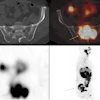
 A 74-year-old patient with metastatic prostate cancer with a positive left iliac wing lesion (SUVmax of 35.02) seen on (A) Zr-89 DFO-YS5 PET maximum projection image (MIP) and (B) PET/CT (red arrows). The lesion was graded 5 on PET but was not visible on CT (D) or (D) technitium-99 MDP bone scan. Felicia Tang, MD, and SNMMI
“CD46-targeted PET imaging with Zr-89 DFO-YS5 was well tolerated and demonstrated high tumor uptake in men with mCRPC,” the researchers wrote.
Ongoing clinical trials will compare the detection efficiency of Zr-89 DFO-YS5 to PSMA-PET and further evaluate its utility as a predictive imaging biomarker in patients with mCRPC most likely to respond to CD46-targeted therapy, the group concluded.
Check out AuntMinnie’s full coverage of SNMMI 2026 on our ShowCast.