PET/CT can fulfill an unmet need by helping diagnose cardiovascular complications in chronic kidney disease patients undergoing hemodialysis, researchers have reported.
The finding is from a small prospective pilot study in patients with chronic kidney disease, with F-18 sodium fluoride (NaF) PET/CT scans quantifying changes in cardiac and aortic vascular calcifications, noted lead author Jonathan Dyke, PhD, of Weill Cornell Medicine in New York City, and colleagues.
“Our findings demonstrate that F-18 NaF PET can quantitatively assess vascular calcification activity and ongoing osteogenesis, offering a sensitive tool to characterize mineral metabolism abnormalities in this highly vulnerable group,” the researchers wrote. The study was published on February 7 in BMC Nephrology.
Vascular calcification is the build-up of calcium mineral deposits in artery walls and heart valves, which can lead to stiffening and cause major cardiovascular adverse events. It affects up to 90% of patients undergoing maintenance hemodialysis, the authors explained.
Current monitoring of vascular calcification in hemodialysis patients is not standard of care, and when prescribed, it is typically done by routine x-rays or CT scans, which do not provide metabolic information, they noted. Conversely, F-18 NaF PET/CT can visualize vascular calcification at the molecular level, as the radiotracer binds to areas of active bone mineral formation.
In this study, to assess whether F-18 NaF PET/CT could be useful for monitoring vascular calcification in patients, the researchers recruited seven participants with chronic kidney disease (3 females, 4 males; average age, 65) being treated with hemodialysis. All subjects underwent imaging (Biograph mCT 64, Siemens Healthineers) at baseline and 9.9 months following. They used a Patlak analysis to quantify active mineral deposition based on the rate of radiotracer uptake (Ki).
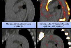
 The image on top shows representative vascular calcifications (VCs) in both the aorta and heart in a F-18 NaF static PET image. The increased PET uptake in the VCs is concordant with increased signal and Hounsfield Unit density on the CT image below. BMC Nephrology
In addition, coronary artery calcium scores derived from the CT scans ranged from moderate to severe in six patients and extensive in one, indicating the presence of atherosclerotic disease, the study authors noted.
“This is the first study to provide evidence that serial F-18 NaF PET imaging is both feasible and informative in this population,” the group wrote.
While this was a pilot study limited by a small sample size, it demonstrates a unique opportunity for a non-invasive imaging technique like F-18 NaF PET/CT to identify vascular calcification in hemodialysis patients, the researchers concluded.
The full study is available here.