PET/CT imaging with gallium-68 (Ga-68) trivehexin (TVR-PET) significantly improves the detection of lesions in patients with primary hyperparathyroidism, according to research presented December 1 at RSNA.
The finding is from a prospective study in 38 patients in whom TRV-PET detected 92% of lesions, compared with 74% with standard technetium-99m sestamibi SPECT (MIBI), according to presenter Dilara Zorba, MD, a resident at Istanbul University in Turkey.
“Our data supports gallium-68 trivehexin PET/CT as a valuable tool in difficult cases such as small lesions,” she said.
Primary hyperparathyroidism (PHPT) is one of the most common endocrine disorders. The most common cause is parathyroid adenomas (noncancerous tumors), and the main treatment is surgery, with accurate preoperative imaging of hyperfunctioning tissue essential for positive outcomes, Zorba explained.
Ga-68 trivehexin is a novel radiotracer that targets Integrin beta-6, a protein expressed by tumors and has shown promise in patients with head and neck squamous cell carcinoma and pancreatic ductal adenocarcinoma, she noted. It was discovered incidentally that it could be useful in patients with PHPT.
“We discovered by chance that it can also demonstrate intense uptake in parathyroid lesions,” Zorba said.
To further evaluate TRV-PET's use in these cases, the researchers recruited 38 patients (age range, 18 to 73 years; 29 women) with biochemically confirmed PHPT. All participants underwent cervical ultrasonography, MIBI, and TRV-PET, with additional imaging including 4D-CT (n = 16) and F-18 choline PET/CT (n = 8). Two nuclear medicine physicians independently evaluated the imaging studies visually and semiquantitatively.
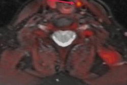
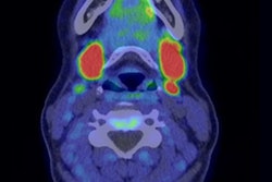
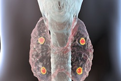
According to a patient-based analysis, TRV-PET detected lesions in 35 of the 38 (92%) patients, while MIBI was positive in 28 (74%) (p < 0.05). In lesion-based analysis, TRV-PET identified 49 of 50 lesions (98%), 34 of which were smaller than 1 cm, while MIBI only detected 29 lesions (58%) (p < 0.01), Zorba reported.
In addition, TRV-PET showed clear uptake in 18 lesions with equivocal MIBI findings and revealed two lesions missed by both MIBI and F-18 choline PET/CT. Only one lesion identified by MIBI was negative on TRV-PET. Finally, TRV-PET successfully localized residual lesions in all seven patients with persistent disease after surgery, Zorba noted.
“[Ga-68 trivehexin PET/CT] showed statistically significantly higher detection rates than MIBI in both patient- and lesion-based analysis,” she said.
Ultimately, the first study of the use of TVR-PET in PHPT was published only last year and included just 13 patients, Zorba noted. This extended data support its use for detecting hyperfunctioning parathyroid tissue when conventional imaging is inconclusive in both initial evaluation of patients and in those with persistent hyperparathyroidism after surgery, she said.
“Our results need to be supported by a larger series,” Zorba concluded.