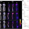
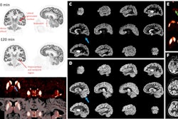
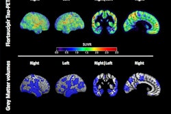
Tau PET brain scans are associated with a high risk of clinical progression in both preclinical and symptomatic stages of Alzheimer’s disease (AD), according to a study published June 16 in JAMA.
The findings are from an analysis of imaging from 6,514 participants from 13 countries and underscore the potential of tau PET as a biomarker for staging the disease, noted lead author Alexis Moscoso, PhD, of the University of Gothenburg in Sweden, and colleagues.
“A positive tau PET scan occurred at a nonnegligible rate among cognitively unimpaired individuals, and the combination of [amyloid] PET positivity and tau PET positivity was associated with a high risk of clinical progression in both preclinical and symptomatic stages of AD,” the group wrote.
Beta-amyloid plaque deposits and tau protein neurofibrillary tangles are two hallmarks of Alzheimer’s disease. PET scans are effective methods for detecting these pathologies, yet the consequences of positive amyloid PET scans in the early stages of the disease remain uncertain. This is because amyloid positivity can occur in older individuals who remain symptom-free over their lifetime, the authors explained.
Similarly, previous studies on tau PET have relied on the use of varying, nonclinically applicable definitions of tau PET positivity, leading to conflicting results on crucial metrics that inform its potential utility, the group explained.
Thus, in this study, the researchers noted that they used the only biomarker of neurofibrillary tangles approved for clinical use by the U.S. Food and Drug Administration (FDA) and the European Medicines Agency (EMA), namely the imaging agent flortaucipir (Tauvid, Eli Lilly).
The study included data pooled from 6,514 participants from 13 countries collected between January 2013 and June 2024. The main outcome of the analysis was the frequency of tau PET positivity and absolute risk of clinical progression. Among participants (mean age, 69.5 years old; 50.5% female), the median follow-up time ranged from 1.5 to 4 years.
According to the analysis, out of 3,487 cognitively unimpaired participants, 349 (9.8%) were tau PET positive. The estimated frequency of tau PET positivity was less than 1% in those younger than 50, and increased from 3% at 60 years old to 19% at 90 years old. Tau PET positivity frequency increased across mild cognitive impairment (MCI) by 43% and AD dementia by 79% at 75 years old.
In addition, most tau PET-positive individuals (92%) were also amyloid PET-positive. Cognitively unimpaired participants who were positive for both amyloid PET and tau PET had a 57% risk of progression to MCI or dementia over the following five years compared with both amyloid PET-positive/tau PET-negative (17%) and amyloid PET-negative/tau PET-negative (6%) individuals.
Finally, among participants with MCI at the time of the tau PET scan, an amyloid PET-positive/tau PET-positive profile was associated with a 70% absolute risk of progression to dementia after five years, the researchers reported.
“Tau PET positivity is detectable in a small but nonnegligible proportion of individuals in the preclinical stages of AD, with increasing frequency in symptomatic stages of the disease,” the group wrote.
Ultimately, the research contributes to a better understanding of the potential of tau PET in clinical settings, the authors concluded.
The full study is available here.


