T1/T2-weighted ratio imaging via MRI-guided focused ultrasound (MRgFUS) improves the lesion normalized contrast of chronic brain lesions, according to research published December 16 in Radiology.
A team led by PhD candidate Begoña Andikoetxea from the Clínica Universidad de Navarra in Pamplona, Spain, reported that T1/T2-weighted ratio imaging substantially improved chronic lesion contrast versus T2-weighted imaging. It also reduced interobserver variability and measurement error.
“These findings support the utility of T1/T2-weighted ratio imaging as a valuable tool for assessing [MRgFUS imaged] chronic lesions, with potential applications in clinical practice and research,” the Andikoetxea team wrote.
MRgFUS thalamotomy can treat tremor disorders in patients with either essential tremor or tremor-dominant Parkinson's disease. T2-weighted imaging is typically used to evaluate thalamic lesions identified by MRgFUS. However, T2-weighted imaging has variable lesion visibility during follow-up in some patients. This is due to lesions showing low contrast or becoming undetectable, the researchers wrote.
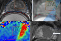
 Definition of regions for lesion normalized contrast calculation on T2-weighted images. (A) Axial T2-weighted image at the 6-month follow-up shows the lesion (outlined in red) in a patient who underwent left-sided MRI-guided focused ultrasound thalamotomy. (B) The corresponding baseline T2-weighted image with a cylindrical reference region (red circle, 16-mm diameter, matching the lesion section number and centered at the lesion centroid) used to measure normal tissue intensity. The same regions were used for lesion normalized contrast calculation on T1/T2-weighted ratio images.RSNA
T1/T2-weighted ratio imaging stems from the mathematical division of T1-weighted by T2-weighted images with additional preprocessing. This approach aims to improve contrast in myelin-rich brain structures, which can help evaluate tissue integrity and microstructural changes in neurodegenerative diseases.
Andikoetxea and colleagues studied whether T1/T2-weighted ratio imaging improves the lesion-normalized contrast of chronic MRgFUS thalamotomy lesions. They compared the results with those of T2-weighted imaging.
The retrospective study included 54 patients, including 27 with essential tremor and 27 with tremor-dominant Parkinson's disease.
T1/T2-weighted ratio images led to higher lesion normalized contrast, including for patients with tremor recurrence.
Comparison between T1/T2-weighted ratio images, T2-weighted images | |||
Measure | T2-weighted images | T1/T2-weighted ratio images | p-value |
Lesion normalized contrast | 19.2% | 36.3% | < 0.001 |
Normalized contrast for patients with tremor recurrence | 25.3% | 45.2% | 0.03 |
Between-evaluator agreement | 0.81 | 0.94 | < 0.05 |
The researchers noted that the improvement in between-evaluator agreement is a critical finding that suggests lower variability.
“This improvement facilitates the more accurate detection of insufficient lesioning and supports informed clinical and surgical decisions, such as re-treatment in patients with tremor recurrence, especially those with low-visibility T2-weighted lesion,” they wrote.
Further analysis showed a positive correlation between lesion overlap with the ventrolateral posterior ventral thalamic nucleus -- the part of the brain that receives information for pain, temperature, and touch -- and tremor relief for both imaging modalities.
The study authors called for future studies to explore the use of T1/T2-weighted imaging against that of other advanced MRI modalities to further improve chronic lesion assessment.
Read the full study here.